|
|
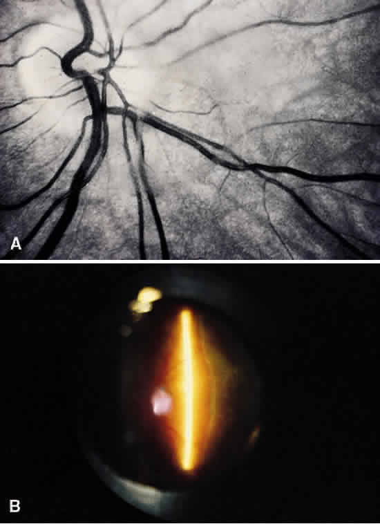
| Fig. 17. Posterior vitreous detachment. A: Fundus photograph of posterior vitreous detachment demonstrates the prepapillary hole in the posterior vitreous cortex seen anterior to the optic disc, which is slightly below and to the right of the optic disc in this photograph. B: Preset lens biomicroscopic photograph of PVD shows a slit beam illuminating the retina and optic disc in the center of this photograph. To the right is the detached vitreous. The posterior vitreous cortex can be seen as the dense, whitish gray, vertically oriented linear structure to the right of the slit beam. (Courtesy of C. L. Trempe, MD) |