|
|
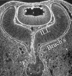
| Fig. 5. Immunohistochemistry of posterior interfaces in the human embryo. This specimen, taken at about the 9-week stage of embryogenesis, was stained with an anti-ABA fluorescent marker that binds to extracellular components of the basal laminae. The continuity of the basal laminae destined to become the internal limiting lamina (ILL) and Bruch's membrane is evident. (Courtesy of Greg Hageman, PhD) |