|
|
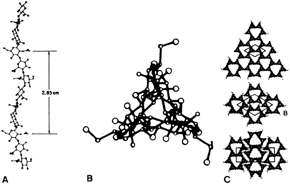
| Fig. 3. Schematic diagram of hyaluronan (HA) molecule (B). A: The left-handed threefold helix is viewed perpendicular to the helix axis and in the center along the helix axis. (From Swann DA. Chemistry and biology of the vitreous. Int Rev Exp Pathol 22:1, 1980). C: Possible packing arrangements for HA molecules viewed along the axis of the helix. ( B and C From Sheehan JK, Atkins EDT, Nieduszynski IA. X-ray diffraction studies on the connective tissue polysaccharides: Two dimensional packing scheme for threefold hyaluronic chains. J Mol Biol 91:153, 1975) |