|
|
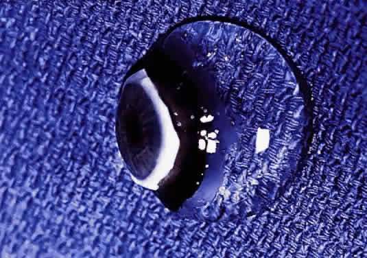
| Fig. 1. Vitreous from a 9-month-old child. The sclera, choroid, and retina were dissected off the vitreous, which remains attached to the anterior segment. Because of the young age of the donor, the vitreous is almost entirely gel. Thus, the structure is solid and maintains its shape, although situated on a surgical towel exposed to room air. A band of gray tissue can be seen posterior to the ora serrata. This is peripheral retina that was firmly adherent to the vitreous base and could not be dissected away without disrupting the vitreous base. (Courtesy of the New England Eye Bank, Boston, MA) |