|
|
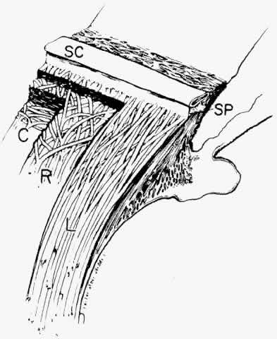
| Fig. 35. Diagram of ciliary muscle divisions: circular (C), radial (R), and longitudinal (L). Anterior attachments to the collagenous scleral spur (SP) and the trabecular beams are indicated. SC, Schlemm's canal. (Modified from Hogan MJ, Alvarado JA, Weddell JE: Histology of the Human Eye. Philadelphia, WB Saunders, 1971; and Rohen JW: Der Ziliarkorper als functionelles System. Morph Jahrbuch 92:415, 1952) |