|
|
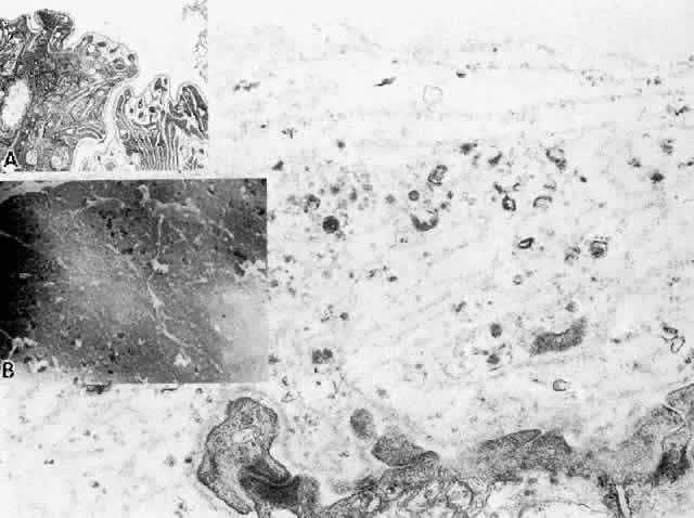
| Fig. 17. Changes in the ciliary nonpigmented epithelial basement membrane that occur with aging.Main figure shows multilaminar pattern of basement membrane proliferation filled with vesicles and granular debris at age 20. (X 54,600) Inset A. Thin basement membrane with patchy multilaminar change at age 7. (X 15,000) Inset B. Scanning electron micrograph showing fibrogranular mossy appearance of basement membrane surface over sides of anterior ciliary processes at age 20. (X 10,200) |