|
|
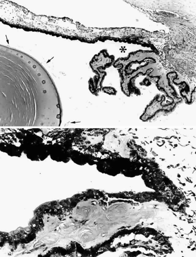
| Fig. 7. The ciliary sulcus. A. Cross section through a ciliary sulcus (asterisk) of a 61-year-old patient. Arrows indicate zonular fibers. Lens is on left. B. Irregular iris pigment (above) and ciliary nonpigment epithelial cells line the sulcus. (hematoxylin-eosin, A, X 43; B, X 165) |