|
|
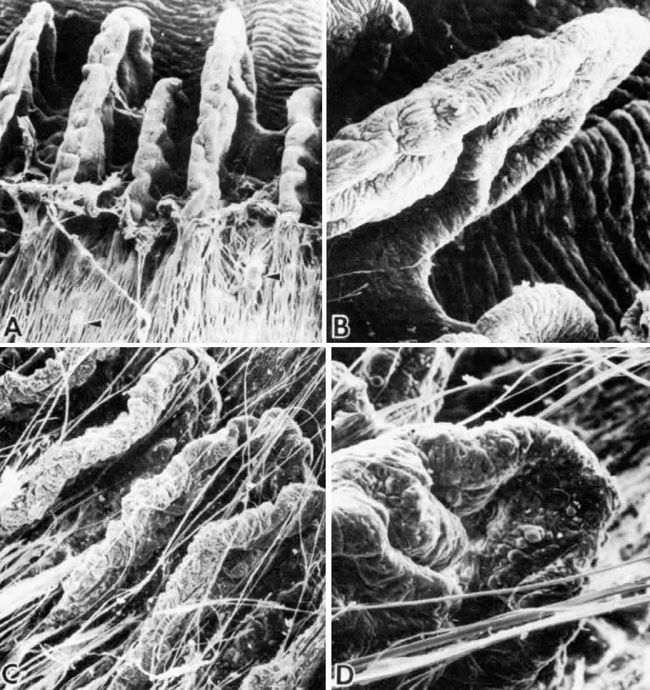
| Fig. 6. Scanning electron microscopy of the ciliary processes of an infant and adult. A and B. Age 3 months. The zonules have been rolled back to reveal the simple infantile processes. Several minor plicae (arrowheads) are visible. The peripheral iris folds are seen above. (A, X 60; B, X 115) C and D. Age 85 years. The processes are larger, more convoluted, and warty. (C, X 45; D, X 110) |