|
|
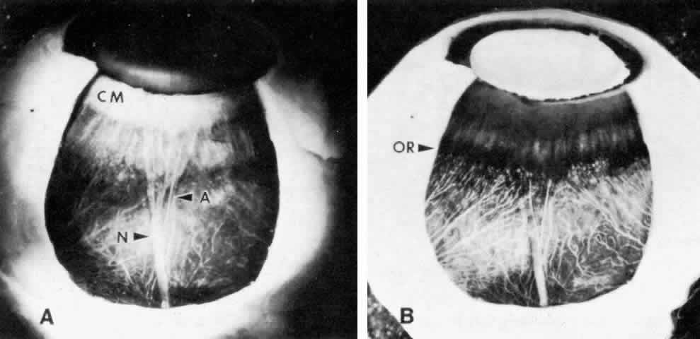
| Fig. 5. Outer surface of ciliary body seen after removal of a scleral window, cornea, and iris. A. Most of the ciliary muscle (CM) lies in the anterior half as a white band. A flat ribbon-like long posterior ciliary nerve (N) and thin artery (A) bifurcate shortly before the ora serrata. B. Transillumination indicates the actual site of the ora serrata and relationships to the choroidal vessels. |