|
|
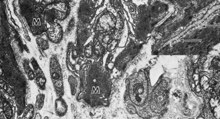
| Fig. 18. Sphincter muscle (M) cells are surrounded by basement membrane and show patches of electron-dense material and myofilaments. Synaptic vesicles are indicated (asterisks) (×23,100). Inset shows smooth muscle cell with pinocytotic vesicles (arrows) adjacent to the plasma membrane (×23,100). |