|
|
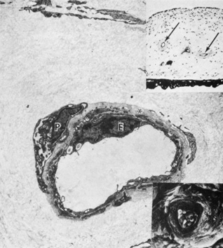
| Fig. 16. Upper inset shows small iris stromal arteries (arrows) (Masson trichrome, ×130). Scanning electron micrograph in lower inset shows red blood cells in the lumen of an iris stromal artery surrounded by a wide collagenous zone (×800). The transmission electron micrograph shows an endothelial (E) lining of the stromal blood vessel with adjacent basement membrane (asterisk). A pericyte is indicated by P (×6450). |