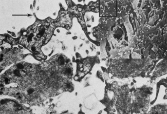
Fig. 10.
Microvilli
(arrow)
are present in the fibroblasts of the anterior border layer (×18,000).
Inset
shows numerous microvilli
(box)
on the cell surface of fibroblasts (scanning electron microscopy, ×4800).