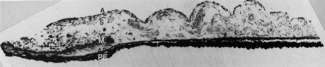
Fig. 5.
The histology of the iris may be divided into five layers: anterior border
(A),
stroma
(S)
, muscle
(M) ,
anterior pigment epithelium
(AP) ,
and osterior pigment epithelium
(PE)
(hematoxylin-eosin, ×63).