|
|
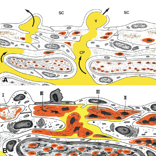
| Fig. 33. Schematic drawings of the cribriform layer in normal (A) and glaucomatous eyes (B). Aqueous pathways (CP) are obstructed by plaque material deposited within the subendothelial region. Arrows indicate direction of flow. E, endothelium of Schlemm's canal; SC, Schlemm's canal; V, giant vacuole; I, II, III, type I, II, and III plaques. |