|
|
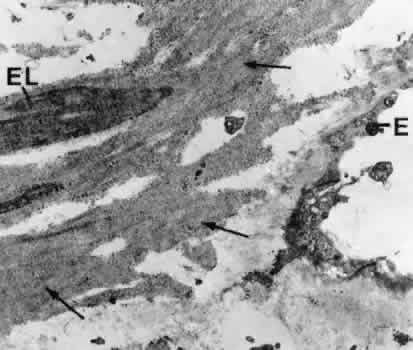
| Fig. 29. Electron micrograph of a tangential section through the cribriform layer of the trabecular meshwork in a case of primary open-angle glaucoma (× 7,500). Note that the plaques have developed from the sheaths (arrows) of the elastic-like fibers (EL) in the subendothelial region. E, trabecular endothelial cell. |