|
|
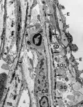
| Fig. 28. Electron micrograph of the cribriform layer of the trabecular meshwork in a 50-year old patient (× 9,000). The subendothelial net of elastic-like fibers (arrows) and its connection with the fiber system of the corneoscleral lamellae (arrowheads) can be recognized. E, endothelium of Schlemm's canal; CL, cribriform layer cell; EL, elastic-like fiber cell; SD, sheath-derived plaques; TB, trabecular lamellae. ( Lütjen-Drecoll E, Rohen JW: Morphology of aqueous outflow pathways in normal and glaucomatous eyes. In Ritch R, Shields MB, Krupin T [eds]: The Glaucomas, vol 1, pp 41–74. St. Louis, CV Mosby, 1989) |