|
|
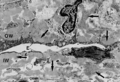
| Fig. 27. Electron micrograph of inner wall (IW) and outer wall (OW) of Schlemm's canal in a case of primary open-angle glaucoma (× 17,280). Note the accumulation of sheath-derived plaque material (arrows), deposited both in the inner and outer wall region. E, endothelium of Schlemm's canal. ( Lütjen-Drecoll E, Rohen JW: Morphology of aqueous outflow pathways in normal and glaucomatous eyes. In Ritch R, Shields MB, Krupin T [eds]: The Glaucomas, vol 1, pp 41–74. St. Louis, CV Mosby, 1989) |