|
|
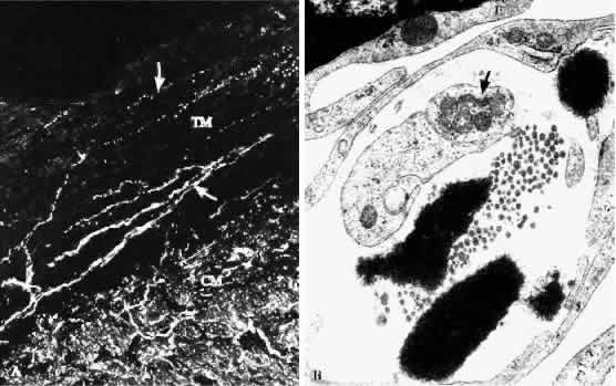
| Fig. 22. A. Histologic tangential section through the outer corneoscleral trabecular meshwork (TM) and the ciliary muscle tips stained for the panneuronal marker PGP. Note that not only the ciliary muscle (CM) shows intense staining, but that there are also circulary running nerve fibers and terminals in the trabecular meshwork (arrows) (× 380). B. Nerve terminals containing numerous mitochondria (arrow) are present beneath the endothelial lining of Schlemm's canal (E) and are in direct contact with extracellular matrix components. (Electron micrograph, × 24,000) |