|
|
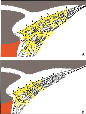
| Fig. 20. Trabecular meshwork in two different functional stages. A. After ciliary muscle contraction induced by pilocarpine treatment. The trabecular meshwork is expanded; the scleral spur has been moved posteriorly. The entire filtration area is working (arrows). B. After reduction of ciliary muscle tone (e.g., after atropine treatment). The anterior parts of the trabecular meshwork are collapsed so that pathways to the endothelial lining are blocked (x). (Rohen JW: The evolution of the primate eye in relation to the problem of glaucoma. In Lütjen-Drecoll E [ed]: Basic Aspects of Glaucoma Research, vol 1. Stuttgart, Schattauer Verlag, 1982) |