|
|
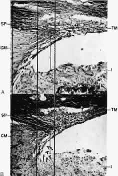
| Fig. 19. Light micrographs of sagittal sections through the chamber angle of an eye enucleated because of a choroidal melanoma in a 57-year-old patient (× 180). The eye was sagittally halved; one half was treated with atropine (A) and the other half with pilocarpine (B). Note the posterior movement of the scleral spur (SP) and the spreading of the trabecular meshwork (TM) after ciliary muscle (CM) contraction. I, iris. |