|
|
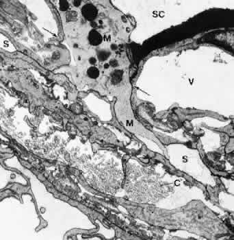
| Fig. 17. Electron micrograph of a sagittal section through the inner wall of Schlemm's canal (SC) after anterior chamber perfusion with cationized ferritin (cynomolgus monkey, × 21,800). Note the large macrophage (M) squeezing through the intercellular space (arrows) of the inner wall endothelium of Schlemm's canal (SC). C, collagenous fibers; V, giant vacuole; S, subendothelial space. |