|
|
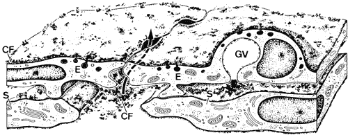
| Fig. 16. Structure of the inner wall endothelium (E) of Schlemm's canal (SC) showing a transcellular pathway through a giant vacuole (GV) and a paracellular route (arrow) labeled with cationized ferritin (CF). S, subendothelial cells. (Epstein DL, Rohen JW: Morphology of the trabecular meshwork and inner wall endothelium after cationized ferritin perfusion in the monkey eye. Invest Ophthalmol Vis Sci 32:160, 1991) |