|
|
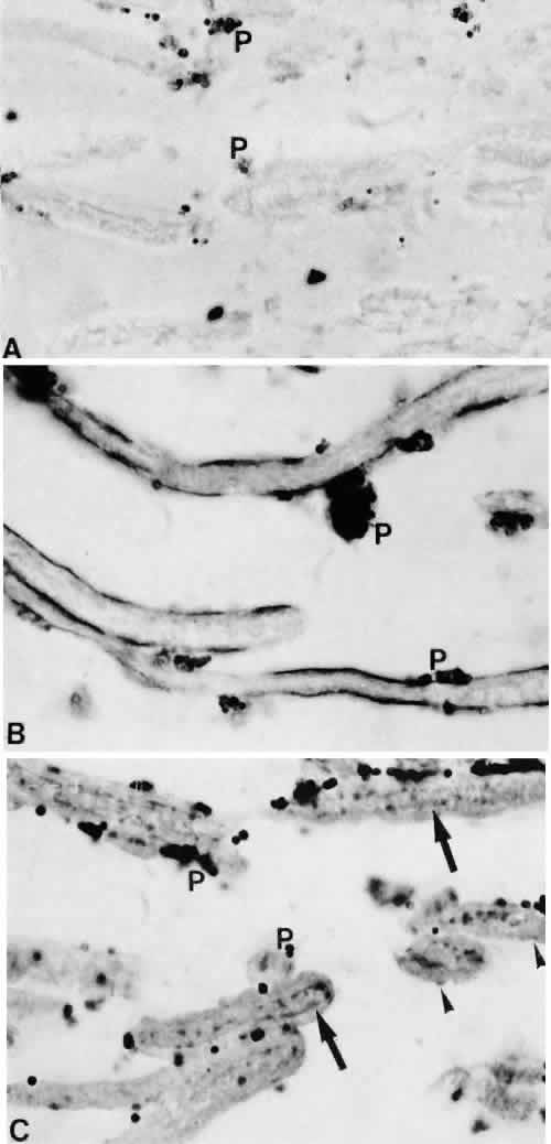
| Fig. 11. Immunohistochemical staining for type IV and VI collagen. Sagittal sections through the trabecular meshwork of a normal eye of a 65-year-old man (× 1,300). In all three sections, goat anti-rabbit IgG, conjugated with colloidal gold (5 nm), was used as secondary antibody for 60 minutes. A. Control section incubated with nonimmunized serum; the dark spots are pigment granules (P) in the trabecular endothelial cells. B. After incubation with antibodies against type IV collagen. Note the dark-stained line underneath the endothelium in the region of the basement membrane. The central core of the lamellae is almost completely unstained. C. After incubation with antibodies against type VI collagen. Within the central core of the lamellae an intense staining is seen where the elastic-like fibers are located (arrows). In the basement membrane, dark-stained spots are visible (arrowheads). ( Lütjen-Drecoll E, Rittig M, Rauterberg J et al: Immunomicroscopical study of type VI collagen in the trabecular meshwork of normal and glaucomatous eyes. Exp Eye Res 48:139, 1989) |