|
|
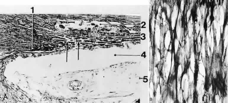
| Fig. 5. Light micrographs of the human trabecular meshwork.A. Sagittal section (orcein stain, × 150). 1, scleral spur; 2,Schlemm's canal; 3, trabecular meshwork; 4, chamber angle; 5, iris. B. Tangential section (plane of sectioning indicated in A by arrows); silver impregnation after Gomori (× 450). Note the regular network of collageneous fiber bundles. (Modified from Rohen JW, Unger HH: Zur Morphologie und Pathologie der Kammerbucht des Auges. Wiesbaden, Steiner Verlag, 1959) |