|
|
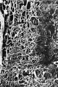
| Fig. 4. Scanning electron micrograph of the human trabecular meshwork. Internal aspect from the chamber angle side (× 1,640). CE, corneal endothelium; IS, iridial strands (remnants of pectinate ligament); U, uveal meshwork; arrows, corneoscleral meshwork. (Modified from Lütjen-Drecoll E, Rohen JW: Morphology of aqueous outflow pathways in normal and glaucomatous eyes. In Ritch B, Shields MB, Krupin T (eds): The Glaucomas, vol 1, pp 41–74. St. Louis, CV Mosby, 1989) |