|
|
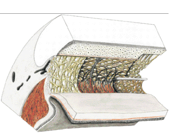
| Fig. 3. Architecture of the trabecular meshwork in the human eye.1, Ciliary muscle; 2, sclera; 3, col-lector channel; 4, Schlemm's canal;5, cornea; 6, iris root; 7, iridialstrands; 8, uveal portion of the trabecular meshwork; 9, corneal endothelium; 10, Schwalbe's line;11, anterior ciliary muscle tendons;12, corneoscleral portion of the tra-becular meshwork; 13, scleral spur; 14, cribriform layer. (Modified from Rohen JW, Unger HH: Zur Morphologie und Pathologie der Kammerbucht des Auges. Wiesbaden, Steiner Verlag, 1959) |