|
|
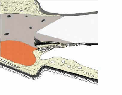
| Fig. 1. Organization of the chamber angle and the trabecular meshwork. A, Nonfiltering portion of the trabecular meshwork; B, filtering portion of the trabecular meshwork; 1, iridial meshwork (pectinate ligament); 2, uveal meshwork; 3, corneoscleral meshwork; 4, cribriform layer (juxtacanalicular or endothelial meshwork); 5, ciliary meshwork (ciliary body band). |