|
|
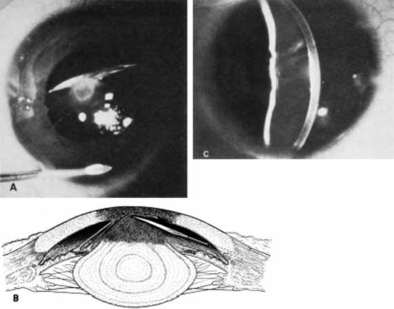
| Fig. 21 Surgical treatment of congenital corneolenticular adhesion. A. Knife needle separates lens stalk from overlying corneal opacity. Note anterior chamber irrigation tube. B. Corneolenticular adhesion, keratoiridial adhesions, and knife needle separating lens from cornea. C. Slit lamp photograph taken 8 years after original surgery shows healed posterior corneal defect with slight scarring. (Waring GO, Parks MM: Successful lens removal in congenital corneolenticular adhesion (Peters anomaly). Am J Ophthalmol 83:526, 1977) |