|
|
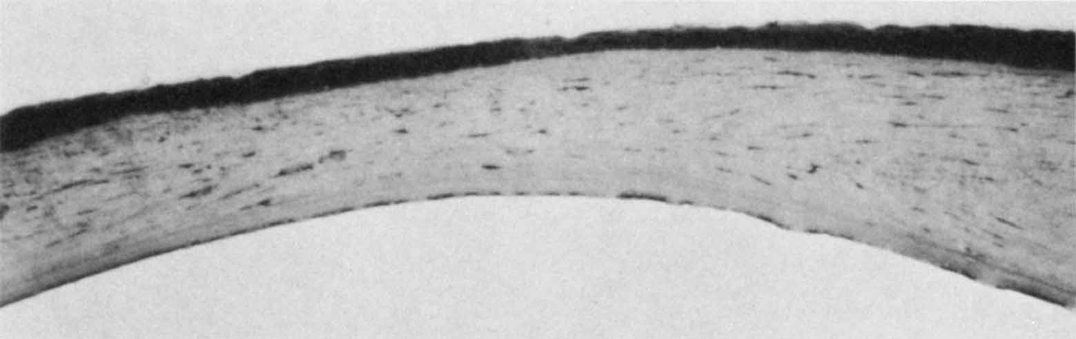
| Fig. 17 Histopathology of central posterior keratoconus. The corneal stroma is thinned centrally. Descemet's membrane is intact but shows abnormal lamellar organization when examined by electron microscopy, and there are few endothelial cells (Toluidine blue, ×60). |