|
|
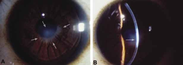
| Fig. 16 Posterior keratoconus. A. Frontal view shows central nebular corneal opacity overlying the margin if the pupil (arrows). B. Slit lamp view of the same cornea shows slight focal posterior indentation (arrow) with overlying full-thickness stromal opacity. |