|
|
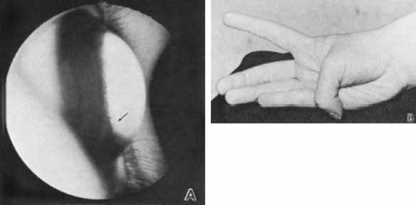
| Fig. 4 Keratoglobus. A. Lateral view demonstrates anterior protrusion of thin cornea. Iris process in the angle are visible (arrow) without gonioscopic lens. B. Increased flexion of thumb emphasizes association with lax joints. Keratoglobus is sometimes classified as Ehlers-Danlos syndrome type VIA. |