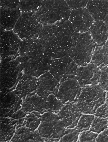
Fig. 21.
Scanning electron micrograph of corneal endothelium. Note the regular hexagonal arrangement of the cells (1,170×). (Courtesy of Drs. Rodrigues, Waring, Hackett, and Donohoo.)