|
|
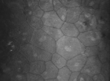
| Fig. 20. Confocal microscopic transverse image of the human corneal endothelium in vivo after penetrating keratoplasty. Note cellular enlargement, polymegathism, and polymorphism. Cell nuclei are clearly visible. Normally non-dividing, endothelial cells enlarge slowly with age to compensate for cell loss, maintaining a continuous lining on Descemet's membrane. Cataract surgery and transplants generally exhibit cell loss. Below about 400 cells per mm2 endothelial decompensation can occur with ensuing edema (500×). (Courtesy of Nidek Technologies.) |