|
|
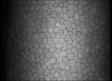
| Fig. 19. Confocal microscopic transverse image of the human corneal endothelium in vivo. In the young normal cornea, the majority of the cells will have a hexagonal outline and they will be fairly uniform in size. The dark spots near the center in many of the cells may represent the central endothelial cilium (500×). (Courtesy of Nidek Technologies.) |