|
|
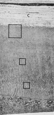
| Fig. 17. Full-thickness view of Descemet's membrane. Stromal collagen (C) with a keratocyte (F) is seen at top. Endothelium (E) is seen at bottom. Large box indicates anterior banded zone of 100-nm spaced collagen. Smaller boxes indicate occasional foci in the amorphous posterior unbanded zone. Arrows point to vesicles on the endothelial basal membrane. (Courtesy of Drs. Rodrigues, Waring, Hackett, and Donohoo.) |