|
|
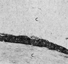
| Fig. 14. Stromal collagen fibrils (c) in uniform spatial arrangement within orthogonal lamellae. A portion of a keratocyte is seen within the interlamellar space. Granular material (asterisk) is visible adjacent to the cell body (30,000×). (Courtesy of Drs. Rodrigues, Waring, Hackett, and Donohoo.) |