|
|
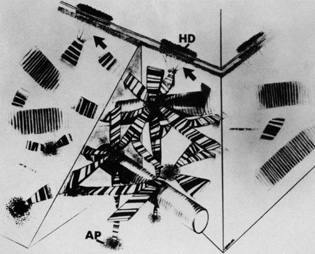
| Fig. 11. Schematic representation of the anchoring fibril network. Separate collagen filaments emerge from the lamina densa, coalesce into banded anchoring fibrils (arrows), and travel distally to insert into anchoring plaques (AP). Note the anchoring filaments between the lamina densa and the hemidesmosomes (HD) with a dense plate feature lying across the filaments within the lamina lucida. Large type I collagen fibrils are interwoven with the anchoring fibril network. (From Gipson IK, Spurr-Michaud SJ, Tisdale AS: Anchoring fibrils form a complex network in human and rabbit cornea. Invest Ophthalmol Vis Sci 28:212, 1987.) |