|
|
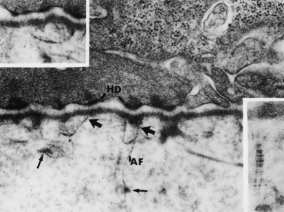
| Fig. 10. Transmission electron micrograph of the epithelial basal lamina in rabbit cornea. Large micrograph shows anchoring filaments within the lamina lucida, oriented between the hemidesmosomes of the basal epithelial cells (HD) and the lamina densa. Anchoring fibrils (AF) travel distally (large arrows) from the lamina densa to insert into electron-dense anchoring plaques (small arrows) (73,900×). Inset right: Cross-banding on the anchoring fibril. Inset left: An anchoring fibril inserting into two adjacent lamina densa sites. (From Gipson IK, Spurr-Michaud SJ, Tisdale AS: Anchoring fibrils form a complex network in human and rabbit cornea. Invest Ophthalmol Vis Sci 28:212, 1987.) |