|
|
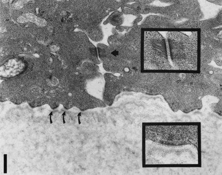
| Fig. 9. Transmission electron micrograph of epithelial basal lamina in the human cornea, Bowman's layer is seen below, with basal epithelial cells above. Note the desmosome junction seen centrally between two basal cells (arrowhead), Magnified view appears in upper right box. Multiple hemidesmosomes are seen along the basal lamina (curved arrows) with platelike features seen within the lamina lucida. Magnified view of a hemidesmosome is seen in the box, lower right, Bar = 0.5 μm. (Courtesy of Roger Beuerman, Ph.D., New Orleans, Louisiana.) |