|
|
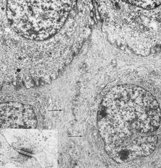
| Fig. 7. Transmission electron micrograph of elaborate interdigitations between epithelial wing cell membranes. Desmosomes are seen along the cell walls, as well as intracellular tonofilaments (arrow). Intracellular rough endoplasmic reticulum, scattered mitochondria, and ribosomes are seen in addition to the prominent nuclei (13,800×). Inset: Desmosome junction (49,500×). (Courtesy of Drs. Rodrigues, Waring, Hackett, and Donohoo.) |