|
|
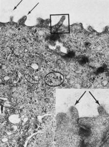
| Fig. 6. Transmission electron micrograph showing the apical membrane of the surface epithelium and intercellular junctions along the lateral membranes. Arrows indicate fine, branching filaments of the glycocalyx emerging from the surface microvilli. A tight junction (box) and densely staining desmosomes are seen along the lateral membranes. Glycogen is evident (circle), as well as numerous vesicles (23,000×). Inset: Arrows point to microvilli from two adjacent surface cells (64,500×). (Courtesy of Drs. Rodrigues, Waring, Hackett, and Donohoo.) |