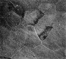
Fig. 5.
Scanning electron micrograph of the anterior surface of the corneal epithelium. Note that the darker cells have fewer microvilli or microplicae near the cell margins (1,500×). (Courtesy of Drs. Rodrigues, Waring, Hackett, and Donohoo.)