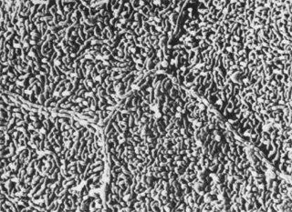
Fig. 3.
Scanning electron micrograph of corneal epithelium surface. Note the numerous microvilli and the cellular margins (12,900×).(Courtesy of Drs. Rodrigues, Waring, Hackett, and Donohoo.)