|
|
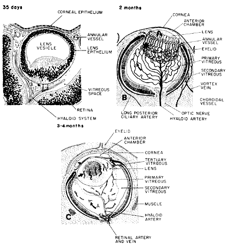
| Fig. 36. Schema of the main features in vitreous development and regression of the hyaloid system shown in drawings of sagittal sections. A. At 5 weeks, the hyaloid vessels and their branches, the vasa hyaloidea propria, occupy much of the space between the lens and the neural ectoderm. One capsulopupillary branch (left) approaches the annular vessel. A capillary net joins the capsula perilenticularis fibrosa (curved arrow), which is composed of some ectodermal fibrils associated with vasoformative mesenchyme from the periphery. The ground substance of this primary vitreous is finely fibrillar. B. By 2 months, the vascular primary vitreous reaches its greatest extent. Arborization of the vasa hyaloidea propria (curved arrow) fills most of the retrolental area. It is embedded in collagen fibrils. An avascular secondary vitreous or more finely fibrillar composition forms a narrow zone between the peripheral (outer) branches of the vasa hyaloidea propria and the retina. Thick arrow indicates the posterior vascular capsule of the lens; in front of it, the channels with a palisadelike arrangement are the capsulopupillary vessels. They connect with the annular vessel. Hooked arrow points to the vessel of the pupillary membrane. Drawing is a composite of embryos at 15 to 30 mm. C. During the fourth month, the hyaloid vessels and the vasa hyaloidea propria, together with the tunica vasculosa lentis atrophies progressively, with the smaller peripheral channels regressing first. Large curved arrow points to remnants of involuted vessels of the superficial portion of the vasa hyaloidea propria in the secondary vitreous. The small curved arrow indicates the pupillary membrane (not sketched). The straight arrow points to the remnants of the atrophied capsulopupillary vessels. Zonular fibers (tertiary vitreous) begin to stretch from the growing ciliary region toward the lens capsule. Vessels through the center of the optic nerve connect with the hyaloid artery and vein and send small loops into the retina (open hollow arrow). The drawing is a composite of fetuses at 75 to 110 mm. |