|
|
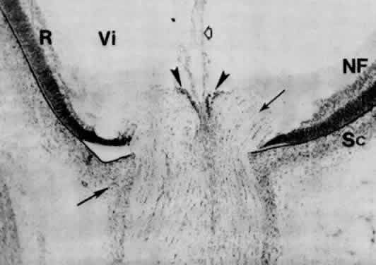
| Fig. 35. Section through the optic nervehead of a 65-mm fetus at 3 months. Bergmeister's papilla (arrowheads) represent the neuroepithelial cells that were displaced toward the center of the optic stalk around the hyaloid artery's entrance at the time (15 mm) when the axons of the ganglion cells made their right angle turn to pass through the stalk. Glial cells of the papilla also extend around the hyaloid artery as its sheath (hollow arrow). Optic nerve fiber bundles surrounded by rudimentary glial septa make a nearly right angle turn toward the scleral foramen (double arrows). Scleral condensation (Sc) merges into that of the developing dura mater (arrow). The inner nuclear and ganglion cell layers end sooner than the outermost cells of the outer nuclear layer of the retina at the exit of the optic nerve fibers. NF, nerve fiber layer; R, retina; Vi, secondary vitreous (× 160). |