|
|
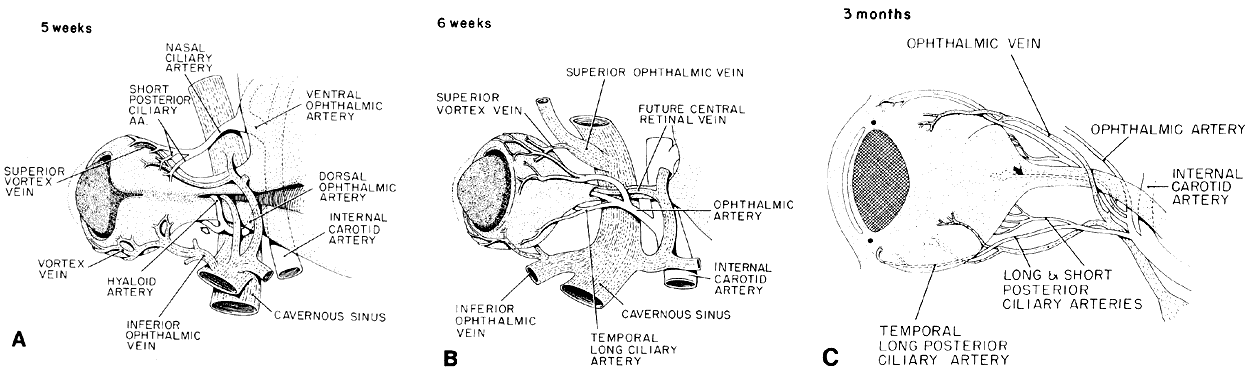
| Fig. 33. Schematic drawing of the optic stalk and early optic nerve formation; their relation to the periocular vasculature. A. Developing optic vesicle and stalk seen from below. Embryonic fissure of the cup is closed except for a notch at the tip. It remains open in the stalk. The hyaloid artery and a terminal branch of the dorsal ophthalmic artery from the internal carotid artery are trapped within the fissure, as is a small twig from the maxillary vein. The other branch of the dorsal ophthalmic artery, which continues outside the cup, is the temporal long ciliary artery (hollow arrow). Ventral ophthalmic artery has a transitory anastomosing branch with the dorsal ophthalmic. The nasal ciliary artery came off this connection, which then disappeared (not shown), so that the dorsal ophthalmic artery eventually remains the only branch from the internal carotid to the eye (see B). Upper and lower venous plexuses draining the blood channels in the mesenchyme next to the pigment epithelium form the primitive superior and inferior vortex veins that connect with the cavernous sinus. B. By 6 weeks, the proximal portion of the fetal fissure is closed up to the small opening for the hyaloid vessels. The interior of the eye is drained by terminal branches of the maxillary vein, which accompany the hyaloid artery and eventually empty into the cavernous sinus. (Redrawn from the film Embryology of the Eye. By permission of the American Academy of Ophthalmology.) C. Relation of the growing optic nerve to the vessels supplying the intraocular structures. The optic nerve has grown to 7 to 8 mm in length and 1.2 mm in width and its orbital portion is being vascularized from the septa (not drawn). Hyaloid artery is marked by curved arrow. (Drawing partly from a cleared specimen at 3 months.) |