|
|
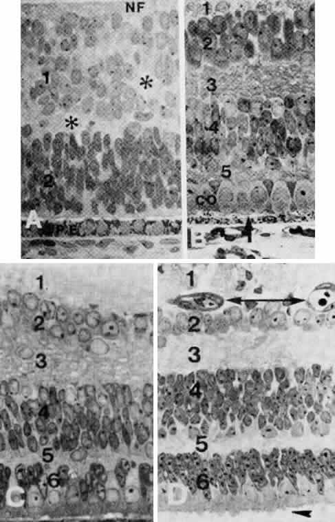
| Fig. 30. A. Portion of retina at the fundus of a fetus at 50 to 55 mm (about 10 weeks). The inner neuroblastic layer (1) is separated from the outer neuroblastic layer (2) by a slowly disappearing layer of Chievitz (*). The pigment epithelium (PE) has a single layer of cells. NF, nerve fiber layer (× 560). B. Portion of the central area from a monkey retina at 76 days, comparable with that of human at approximately 3.5 to 4 months). 1, nerve fiber layer; 2, ganglion cell layer; 3, inner plexiform layer; 4, inner nuclear layer; 5, narrow outer plexiform layer. Cone nuclei (co) are aligned next to the external limiting membrane (arrow) (× 752). C. Section through a portion of the retinal fundus of a macaque fetus at 86 days (comparable with that of human at midterm). 1, nerve fiber layer; 2, ganglion cell layer; 3, inner plexiform layer; 4, inner nuclear layer; 5, outer plexiform layer; 6, outer nuclear layer. D. Section through the fundus of a retina of a fetus at 190 mm (estimated age, 5.5 months). The numbering of the layers is as in C. Double-headed arrow indicates blood vessels in the ganglion cell layer. Arrowhead on the bottom points to photoreceptor inner segments protruding into the extracellular space beyond the external limiting membrane (× 650). (B and C from Smelser GK, Ozanics V, Rayborn M, Sagun D: Retinal synaptogenesis in the primate. Invest Ophthalmol 13:340–361, 1974.) |