|
|
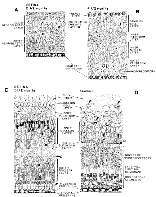
| Fig. 29. Schematic diagram of the developing retina. Region of the posterior pole is represented in sagittal section in every diagram. A. At 2½ months, transient fiber layer of Chievitz, which separated the inner from the outer neuroblastic layers of the primitive retina, is slowly being obliterated by shifting of the nuclear elements and realigning of their processes. Uppermost cells, lying vitread, are differentiating into ganglion cells. Those below the uneven transient layer of Chievitz (*) are immature, but destined to differentiate into amacrine and Müller cells. The future inner plexiform layer will be located between the shifted nuclei of the latter and those of the ganglion cells. The outer neuroblastic layer contains photoreceptor, bipolar and horizontal cell elements. B. At midterm (4½ months), retinal lamination is essentially complete. The ganglion cells have a multilayered arrangement. The inner plexiform layer, composed of fibers of bipolar, ganglion and amacrine cells supported by müllerian fibers, has established sites of primitive conventional and ribbon synapses. In the inner nuclear layer, the still undifferentiated cellular components are recognizable by shape and position. In the outer nuclear layer, large cone nuclei are aligned adjacent to the pigment epithelium and the smaller rod nuclei are positioned more vitread. The outer plexiform layer has primitive lamellar synapses between bipolar cell dendrites and cone pedicles (not indicated). Photoreceptor outer segments are not yet present. C. At 5.5 months, the ganglion cells have thinned out to one to two layers (except in the macular area). The cellular components of the inner nuclear layer include amacrine cells with large pale nuclei in the innermost (vitread) zone of this layer; and pleomorphic, dark-staining müllerian cell nuclei; both these types originally came from the inner neuroblastic layer. Also included are the smaller bipolar cells and large, pale-staining horizontal cells that are in an irregular arrangement sclerad. These two cell types are derived from the outer neuroblastic layer, together with the photoreceptors. The outer plexiform layer has a linear arrangement of synapses between bipolar cells and rod spherules (key symbol). The outer nuclear layer consists of six to seven layers of nuclei; the outermost are cones aligned to the external limiting membrane. Growing photoreceptor outer segments projected into the space between pigment epithelium and external limiting membrane (arrowhead). Cell death is represented by the round dark-centered symbols. D. Newborn retina has the adult configuration with vascularization (arrowheads) reaching the outer limits of the inner nuclear layer. Outer plexiform layer is thinner than that in the adult, but the line of synapses is well established (key symbol). Rod and cone inner and outer segments are fully developed and the tips of the outer segments contact the pigment epithelium. |