|
|
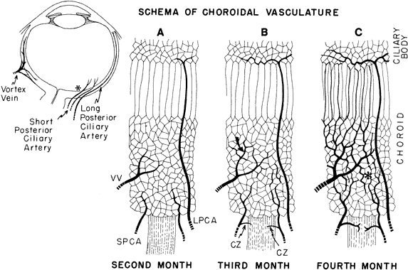
| Fig. 28. Schematic diagram of the development of choroidal vasculature. A. During the second month, primitive vascular meshwork in the mesenchyme around the pigment epithelium connects with small arterial branches of the precursors of the short posterior ciliary arteries (SPCA) that arose from the ophthalmic as two trunks together with the long posterior ciliary arteries (LPCA). These two long arteries run anteriorly through the meshwork, which is drained by tributaries of the infra- and supra-orbital venous plexuses (VV). B. Several future vortex veins (curved arrow) are anterior to the equator of the globe. The two long posterior ciliary arteries bifurcate and start to encircle the region of the future ciliary body. Two to three short posterior ciliary arteries send twigs to the scleral condensation surrounding the optic nerve (CZ). These are the precursors of the circle of Haller-Zinn. In the peripheral choroid the primitive capillary net still has a palisade-like arrangement. C. During the fourth month, a layer of larger vessels form. They are mainly tributaries of the vortex veins (left side). Medium-sized branches of the short posterior ciliary arteries become intercalated between the choriocapillaries and the large venous channels in the posterior choroid (asterisk). Anterior part of the choroidal vasculature, mostly venous, has parallel channels that break into a network in the emerging ciliary region. The long posterior ciliary arteries form the major arterial circle. Interarterial anastomoses are present. (Adapted and redrawn from Heimann K: The development of the choroid in man. Ophthalm Res 3:257, 1972.) |