|
|
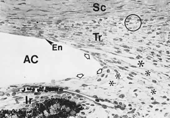
| Fig. 19. Angle at 7 months (approximately 225 mm). Apex of the wedge-shaped trabecular meshwork (Tr) is not in the illustration. The corneal endothelium (En) extends over one third of the trabecular lamellae. The loose tissue in the angle recess is isolated from the anterior chamber (AC) by processes of the reticular and mesenchymal cells (hollow arrows). There are large clefts (*), some of which are confluent, in the angle tissue. The angle recess extends beyond the level of the middle of the trabecular meshwork, and the immature Schlemm's canal (circled) is somewhat behind it. Ir, immature iris; Sc, sclera. (Smelser GK, Ozanics V: The development of the trabecular meshwork in primate eyes. Am J Ophthalmol 71:366, 1971.) |